We Use the Latest Technologies
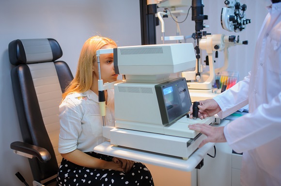
Ocular Coherence Tomography
Optical Coherence Tomography (OCT) provides detailed, cross‑sectional images of the retina, allowing practitioners to view the layers beneath the surface. This imaging can assist in identifying structural changes to retinal nerves and blood vessels that may be associated with conditions such as glaucoma or macular degeneration. These insights help guide clinical decision‑making and monitoring over time..
OCT can also produce high‑resolution images of the front structures of the eye. This may support the early identification and assessment of corneal conditions, including keratoconus and Pellucid Corneal Degeneration, and can be useful in ongoing management and review.
Digital Retinal Imaging
Digital retinal imaging allows us to compare images of the back of your eyes from one visit to the next. This technology assists with early detection of progression or change in the health of your eyes.
Through digital retinal imaging, health concerns, such as Macular Degeneration, glaucoma and retinopathy can be found or monitored.
Medmont Visual Field Screening
Visual field screening allows us to map the sensitivity of the nerves inside your eyes, allowing us to detect any visual field losses due to conditions such as glaucoma or brain injury.
Medmont Corneal Topography
Corneal Topography allows us to map the corneal curvature at the front of your eyes, allowing us to detect any corneal changes due to conditions such as keratoconus or other corneal degeneration. It allows us to measures treatment progress during the corneal reshaping therapy (a.k.a ortho-keratogology), as well as other contact lenses fitting.
Pachymetry
Pachymetry uses a small ultrasound to measure the thickness of the cornea. This is important due to its importance in early detection of the diagnosis of glaucoma.